Tissue Engineering is a relatively new area of endeavor, involving cell
biology and materials science.
It involves the the development of biological materials that can repair,
maintain, or enhance tissue function.
Tissue engineering is an emerging, interdisciplinary field which is full
of promise for those in need of organ and tissue replacement and repair.
To date, the commercial success of tissue engineered products has been
largely unfulfilled.
The development of tissue engineered products is limited by
the lack of laboratory imaging techniques which are capable of
non-destructive imaging of
the three-dimensional morphology as well as the cell response of a tissue
engineering scaffold. In this project, a collinear optical
coherence/confocal fluorescence microscope capable of high-resolution
imaging of porous scaffolds has been developed.
The combined data is then visualized in our interactive, immersive
visualization environment in order to gain new insights into
characteristics that promote cell growth and propagation.
The ability to evaluate cell scaffold viability in this way
will enable the developers of tissue engineered
materials to minimize expensive and time-consuming animal studies.
 |
Papers/Presentations
 |
Judith Terrill, W. L. George , Terrence J. Griffin , John Hagedorn, John T. Kelso, Marc Olano, Adele Peskin , S. Satterfield, James S. Sims, J. W. Bullard, Joy Dunkers, N. S. Martys , Agnes O'Gallagher and Gillian Haemer,
Extending Measurement Science to Interactive Visualization Environments
in Trends in Interactive Visualization: A-State-of-the-Art Survey,
Elena Zudilova-Seinstra, Tony Adriaansen and Robert Van Liere (Ed.)
,
Springer, U.K.,
2009.
Note: Pages: 207-302 |
 |
Judith E. Devaney, Steven G. Satterfield, John G. Hagedorn, John T. Kelso, Adele P. Peskin, William L. George, Terence J. Griffin, Howard K. Hung and Ron D. Kriz, Science at the Speed of Thought,
Lecture Notes in Artificial Intelligence .
Links:
postscript and pdf.
|
 |
J. Hagedorn, J. Dunkers, S. Satterfield, A. Peskin, J. Kelso and J. Terrill, Measurement Tools for the Immersive Visualization Environment: Steps Toward the Virtual Laboratory,
NIST Journal of Research, 112
(5)
,
September-October, 2007,
pp. 257-270.
Links:
htm and pdf.
|
 |
J. Hagedorn, J. Dunkers, A. Peskin, J. Kelso and J. Terrill, Quantitative, Interactive Measurement of Tissue Engineering Scaffold Structure in an Immersive Visualization Environment,
Biomaterials Forum, 28,
2006,
pp. 6-9.
|
 |
J. Hagedorn, J. Dunkers, A. Peskin, J. Kelso and J. Terrill, Quantitative, Interactive Measurement of Tissue Engineering Scaffold Structure in an Immersive Visualization Environemnt,
cover of
Biomaterials Forum, 28,
2006.
|
 |
J. Hagedorn, J. Terrill, J. Kelso and J. Dunkers,
Measurement of Tissue Engineering Scaffolding Material
delivered at DIVERSE Birds of a Feather session at SIGGRAPH 2006, Boston, MA,
August 2, 2006.
|
 |
J. Dunkers, J. Hagedorn, A. Peskin, J. Kelso, J. Terrill and L. Henderson,
Interactive, Quantitative Analysis of Scaffold Structure Using Immersive Visualization
in Proceedings of the 2006 Summer Bioengineering Conference, Amelia Island Plantation Amelia Island, FL,
June 21, 2006.
|
 |
F. A. Landis, M. T. Cicerone, J. A. Cooper, N. R. Washburn and J. P. Dunkers, Developing Metrology for Tissue Engineering: Collinear Optical Coherence and Confocal Fluorescence Microscopies,
IEEE Bioimaging Conference Proceedings,
April 1, 2004.
|
 |
M. T. Cicerone, J. P. Dunkers, N. R. Washburn, F. A. Landis and J. A. Cooper, Optical Coherence Microscopy for in-situ Monitoring of Cell Growth in Scaffold Constructs,
7th World Biomaterials Congress,
2004,
p. 584.
|
 |
J. P. Dunkers, M. T. Cicerone and N. R. Washburn, Collinear Optical Coherence and Confocal Fluorescence Microscopies for Tissue Engineering,
Optics Express, 11
(23)
,
January 1, 2003,
p. 3074.
|
 |
J. P. Dunkers and M. T. Cicerone, Scaffold Structure and Cell Function Through Multimodal Imaging and Quantitative Visualization,
Biomaterials Forum, 25
(3)
,
January 1, 2003,
p. 8.
|
|
|
|
|
 |
|
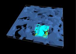
Overview of two scans of a cell scaffold at differing resolutions.
The two scans are registered into the same coordinate system.
|
 |
|
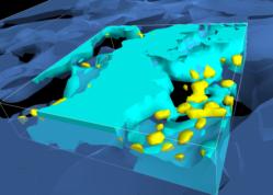
Detailed view of a portion of the registered data sets.
|
 |
|
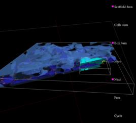
A portion of the immersive display showing several features
of the system: interactive menus, 2D cross-sections,
interactive clipping plane, and transparency.
|
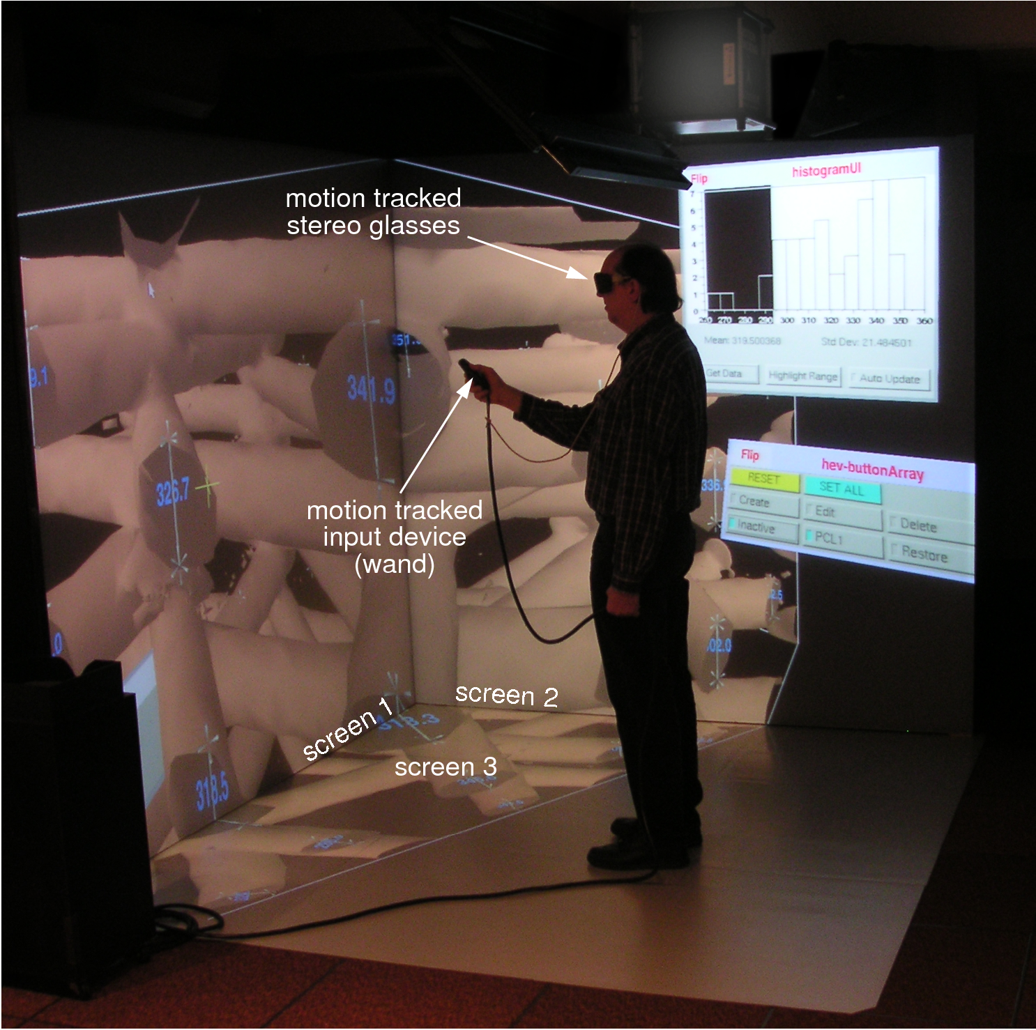 |
| Interactive measurement and analysis in the immersive visualization environment
|
|
|
