The term Body Area Network (BAN) refers to a network intended to be used
in or around the human body.
While this is an emerging field, networks of
medical sensors are anticipated to be a primary application.
Such sensors would either be attached to or implanted in the human body
and would communicate wirelessly both within the body and to
devices outside the body.
For this technology to develop, greater understanding of
radio frequency (RF) propagation through the human body
is needed. In this project, we seek to provide such insight.
This work is assisting the IEEE standards committees to
develop communications standards and will help RF engineers
to design effective communication devices for BANs.
Because experimentation on human subjects is currently not
feasible, RF propagation through the human body is being
modeled in software with a 3D full-wave eletromagnetic
field simulator.
The 3D human body model includes frequency dependent
dielectric properties of 300+ parts in a male human body.
The data produced by this simulation software
is then brought into a 3D immersive visualization system,
which enables researchers to study the modeled RF propagation
through direct interactions with the data.
The simulations are being performed by members of the
Advanced Network Technologies Division and the visualization work
is being done by members of the Scientific Applications
and Visualization Group of the Mathematical and
Computational Sciences Division.
The immersive system includes several important components:
three orthogonal screens that
provide the visual display, the motion tracked stereoscopic
glasses, and a hand-held motion tracked input device.
The screens are large projection video displays that are placed
edge-to-edge in a corner configuration.
These three screens are used
to display a single 3D stereo scene.
The scene is updated based on the position of the user as
determined by the motion tracker. This allows the system to
present to the user a 3D virtual world within which the user can
move and interact with the virtual objects. The main interaction
device is a hand-held three button motion-tracked wand with a
joystick.
This virtual environment allows for more natural
interaction between experts with different backgrounds such as
engineering and medical sciences.
The researchers can look at data
representations at any scale and position, move through data,
change orientation, and control the elements of the virtual
world using a variety of interaction techniques including
measurement and analysis.
For example, we have implemented interactive tools for probing the 3D
data fields. One tool enables the researcher to move the motion-tracked
wand through the virtual scene, yielding a continously updated
display of the value of the data field at the position of the wand.
Another tool enables the user to interactively stretch a line segment through
virtual body, and to generate graphs of the 3D data fields along
that path.
We have found these to be effective tools in getting quantitative
information from the 3D scene and in gaining insight into RF
propagation through the human body.
|
|
|
 |
K. Sayrafian-Pour, W. Yang, J. Hagedorn, J. Terrill and K. Y. Yazdandoost,
A Statistical Path Loss Model for Medical Implant Communication Channels
in
IEEE 2009 Personal, Indoor, and Mobile Radio Communications Symposium
,
September 2009.
Note: Received Best Paper Award. |
|
K. Y. Yazdandoost and K. Sayrafian-Pour,
Channel Model for Body Area Network (BAN)
,
April 2009, Report to the IEEE P802.15.
ID: IEEE 802.15-08-0780-02-0006.
Note: J. Hagedorn, J. Terrill, and W. Yang listed as contributors. |
|
J. Hagedorn, J. Terrill, W. Yang, K. Sayrafian-Pour, K. Y. Yazdandoost and R. Kohno,
A Statistical Path Loss Model for MICS
,
September 2008, Report to the IEEE P802.15.
ID: IEEE 802.15-08-0519-01-0006.
|
|
J. Hagedorn, J. Terrill, W. Yang, K. Sayrafian-Pour, K. Y. Yazdandoost and R. Kohno,
MICS Channel Characteristic: Preliminary Results
,
May 2008, Report to the IEEE P802.15.
ID: IEEE 802.15-08-0351-00-0006.
|
 |
|
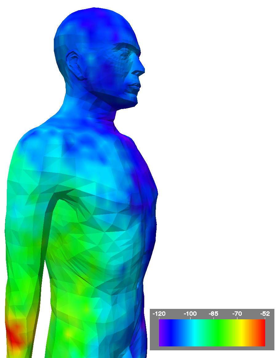
Simulation of a transmitter implanted in the right forearm.
Color denotes signal strength.
|
 |
|
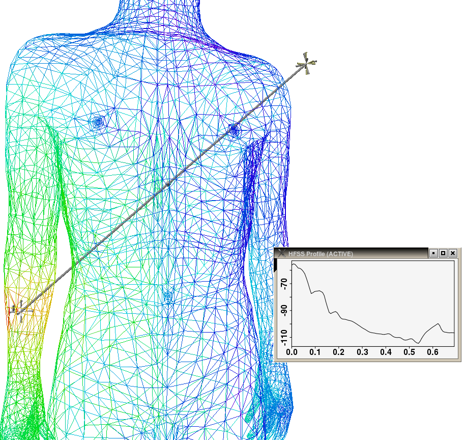
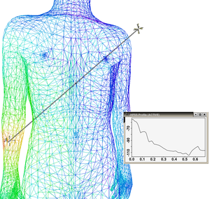
Simulation of a transmitter implanted in the right forearm.
The user has interactively drawn a line segment in the 3D virtual scene.
A graph showing signal strength along that line is displayed.
|
 |
| Video: An Immersive Visualization Environment to Study RF Propagation from Medical Implants
|
|
|